How do we Treat Chronic Dacryocystitis at our centre?
In year 2000, our Chief Surgeon of CSNEH, Dr. Shivendra Sahay became one of the Pioneer Surgeons to demonstrate External DCR surgery with Primary Silastic tube Intubation which had very negligible incidence of failure in management of Chronic Dacryocystitis.
• What is DCR?
Each eye has a fine pipe which drains the tears from the eye. This is a nasolacrimal duct (drain-pipe of the eye). If it gets blocked, the tears and stickiness come out of the eye. The treatment is by dacryocystorhinostomy (DCR). This is a technique by which a new passage is created from the eye into the nose, and the tears can drain out.
• Is it necessary to undergo DCR?
When a nasolacrimal duct is blocked, the dirt and discharge accumulate in the lacrimal sac next to the eye. There is the risk of severe eye infection if the condition is left untreated. There may be swelling, pain, and watering. If a cataract surgery is planned, a blocked nasolacrimal duct increases the risk of dangerous infection; a DCR should be done before the cataract surgery.
• How is DCR performed? What are the outcomes?
DCR can be performed in three ways- externally, through a small (less than half inch) line next to the nose; endonasally- through the nose; and trans-canalicular using Laser DCR. The external DCR leaves a fine mark near the eye; it has the highest success rates, more than 95 out of 100 patients have the problem completely solved after external DCR. An endo nasal DCR is done through the nose, so there is no mark outside. The success rates are a little lower; all nose space inside is not suitable for endo nasal surgery, and it can be done well in selected patients only. Trans-canalicular Laser DCR is a very rapid procedure, with hardly any pain and swelling. However, some of the DCR done with laser may close down again.
• Management of Failed DCR Surgery
As mentioned, about 5 out of 100 patients find that their DCR has closed down again. This may particularly happen in a patient who had multiple attacked of infection earlier, with a history of injury near the nose, or a patient who has frequent nasal allergies and colds. The DCR can be repeated, with addition of silicone intubation to prop the passage open. A typical oculoplastic surgeon will often see patients sent over from elsewhere after the DCR did not work; most such patients can be re-operated successfully by Silastic Tube Intubation which we do in cases of Failed DCR surgeries done elsewhere and referred to us for re-surgery.
• DCR Surgery with Primary Silastic Tube Intubation – Our Treatment of Choice
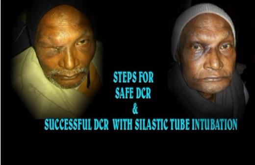
As mentioned above, Dr Shivendra Sahay, our Chief Consultant commonly do DCR surgery with Primary Silastic Tube Intubation. Meaning thereby, we pass a Silicon tube through the ostium (hole) made during the Primary DCR surgery. As a result of which there is very negligible incidence of failure. Also there is no need of patients coming for frequent follow up for painful probing and syringing procedures. The tube is left in its place for 3 to 6 months till formation of permanent bypass channel around the tube. After completion of this period,(3 to 6 months) the tube is gently removed leaving a permanent patent bypass channel for life long. We have presented and published many papers for the above surgery.
OTHER DISEASES ADDRESED IN OCULOPLASTY
2. EYELID RELATED DISEASES
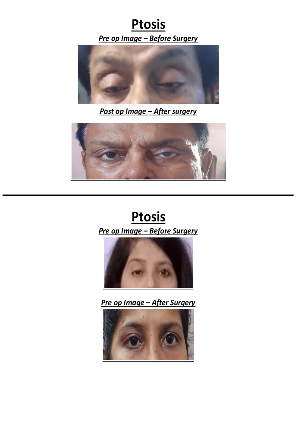
a. Ptosis – Ptosis is drooping of the upper lid. It may be present by birth, or appear later in life. Ptosis is usually corrected by surgery.

b. Entropion – Entropion is in-turning of the eyelid. The eyelashes rub on the eye and cause pain and watering; treatment is by surgery
c. Ectropion – Ectropion is turning outwards of the eyelid margin, causing redness and watering (Link to gallery)Lagophthalmos- where the patient cannot close his eye; this can cause danger to the eye, pain and infection.
d. Chalazion– Chalazion is a condition of localized cheesy Chronic Pus formation in Tarsal gland which appear as well localized round or oval swelling of the upper & lower lid.
e. Eyelid tumors
f. Lid tear repairs with grafts and flaps
g. Blepharoplasty – Correction of eyelid bags
3. ORBIT RELATED DISEASES OFTEN MANAGED HAVING HELP FROM NEURO, ENT & ONCHO SURGEONS
a. Orbital fractures– A fracture in the bones around the eye can cause double vision, and a sunken small appearance of the eye. The fracture is repaired with an implant or plate. The best results are obtained in surgery within 2 weeks of injury, but surgery can also be done later.
b. Thyroid decompression
c. Orbital tumors
4. SOCKET
a. Enucleation/ evisceration– A highly damaged eye may be replacedwith an orbital implant; this restores the comfort and appearance.
b. Socket reconstruction– where the eye has been removed for a serious disease, the socket of the eye may get shrunken. The socket is reconstructed to improve comfort and appearance
c. Customised prosthesis
 0612-2348134
 98014-55691
 csnihospital@rediffmail.com


